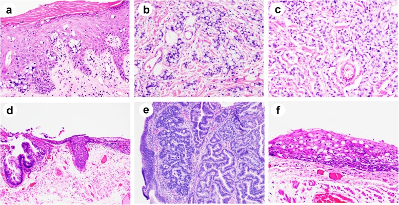
Fig. 2.
Histologic features of secondary perianal Paget’s disease cases associated with underlying malignancy (hematoxylin and eosin). a-c High magnification (× 200) showed intraepithelial Paget’s cells (a) and underlying invasive adenocarcinoma with signet ring cell feature (b) and neuroendocrine feature (c). d Low magnification (× 100) showed intraepithelial Paget’s cells associated with a tubular adenoma with high-grade dysplasia. Higher magnification views (× 200) of (d) showed tubular adenoma with high-grade dysplasia (e) and Paget’s cells (f)

