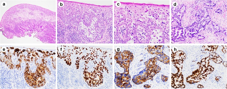
Fig. 3.
Histologic and immunohistochemical features of secondary perianal Paget’s disease with associated invasive adenocarcinoma. a Low magnification showed intraepithelial Paget’s cells and underlying conventional invasive adenocarcinoma. b-d High magnification view of (a) showed Paget’s cells (b, c) and invasive adenocarcinoma (d). Paget’s cells are positive for CK20 (e) and CDX2 (f) by immunohistochemistry. Underlying adenocarcinoma cells are also positive for CK20 (g) and CDX2 (h) by immunohistochemistry. Magnifications: a, × 40; b, × 200; c, × 400; d-h, × 200

