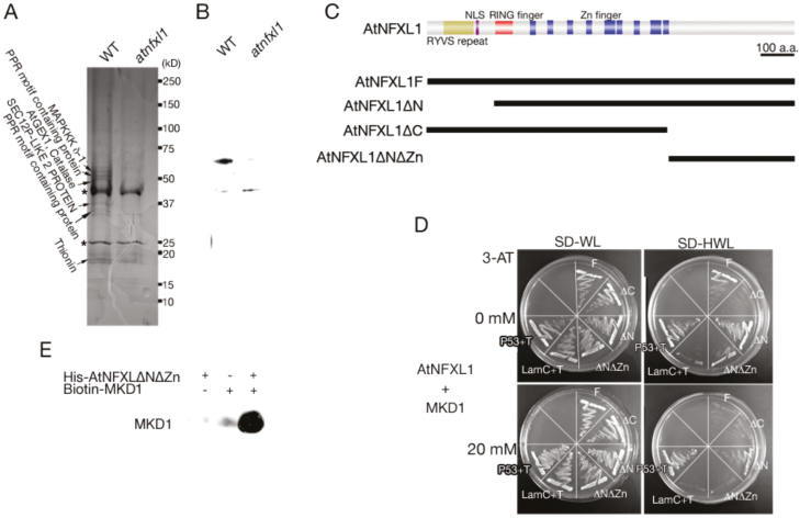
Figure 1.
Protein–protein interaction between MKDI and AtNFXL1. (A) SDS-PAGE of the AtNFXL1 protein-containing complex purified from T-2 toxin-treated WT and atnfxll mutant plants using an anti-AtNFXL1C antibody column. Designations on the left side indicate identified subunits specifically observed in WT. Asterisks indicate non-specific proteins. (B) Western blot analysis of purified AtNFXL1-containing complex using anti-AtNFXL1C antibody. (C) Schematic diagram of full length and partial AtNFXL1 using yeast two-hybrid analysis. (D) The interaction between AtNFXL1 and MKDI was investigated by yeast two-hybrid analysis. The concentrations of 3-amino-1,2,4-triazole (3-AT) are shown on the left. P53+T and LamC+T represent the positive and negative controls, respectively. Similar results were obtained in three independent experiments. (E) The binding of AtNFXL1 protein to the MKD1 protein was examined by pull-down assays. His epitope-tagged AtNFXL1DNDZn protein was applied to a Ni Sepharose High Performance column. Biotin–MKD1 was detected by Transcend™ Non -Radioactive Translation Detection Systems.

