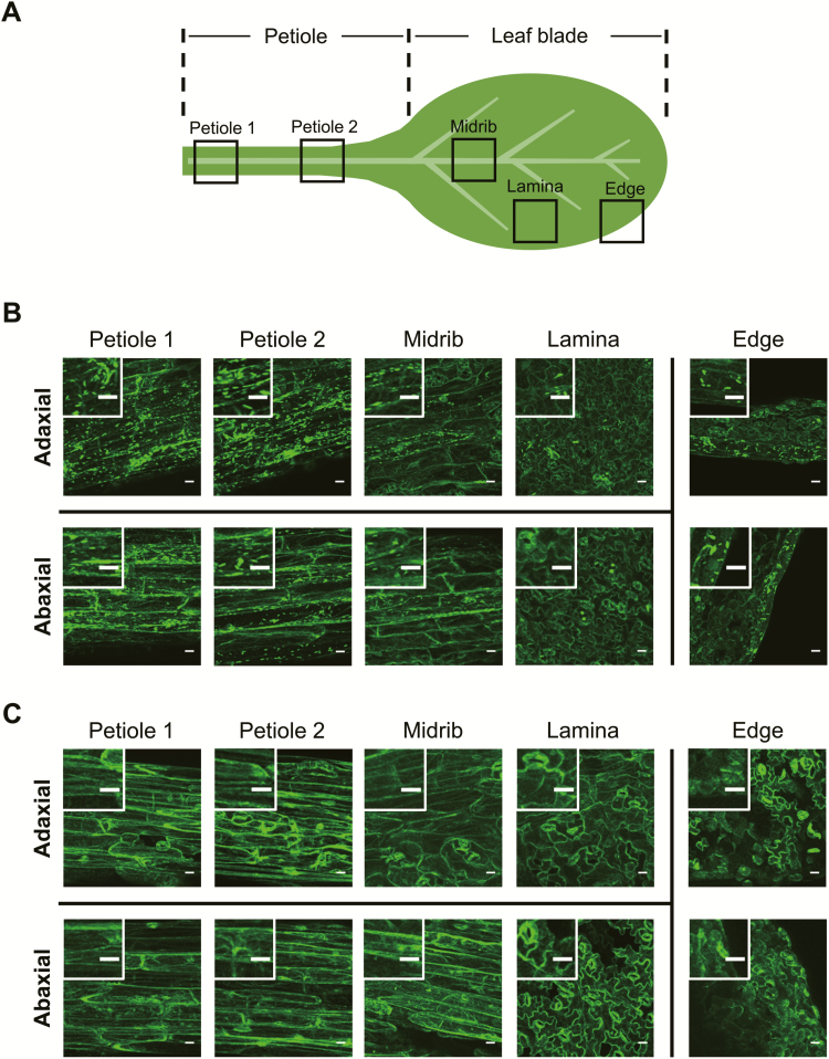
Fig. 2.
Tissue distribution of ER bodies in Arabidopsis leaves. The first true leaf from a 14-day-old plant grown under normal conditions was used. (A) Leaf portions subjected to fluorescence microscopic observation for GFP. (B and C) Representative fluorescence images of different leaf portions from GFP-h (B) and GFP-h nai2-2 plants (C), with enlarged images in the insets. Scale bars=20 μm.

