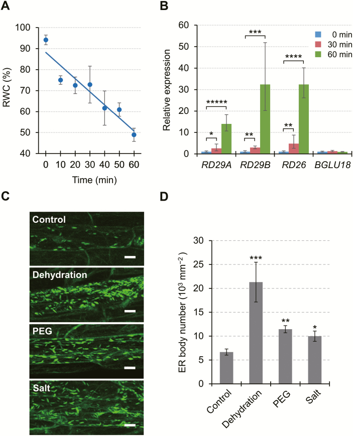
Fig. 3.
Responses of ER bodies to abiotic stress in epidermal cells of Arabidopsis leaf petioles. (A) Effects of drought-induced dehydration stress on leaf relative water content (RWC). Aseptically grown plants were exposed to dry air for up to 60 min by removing the lids from Petri dishes, and leaves were detached for RWC measurements (n=3, where each sample consisted of at least 10 plants). The straight line corresponds to a linear model fitted to the measured data (y= –0.6235x+88.148, r2=0.8959, P<0.005). (B) Effects of dehydration stress on stress-responsive gene expression. Total RNA was extracted from the aerial parts of dehydration-stressed plants at the indicated time points. Transcript levels of target genes were measured by RT–qPCR, normalized to those of SAND as a reference gene, and represented as values relative to the level at the start of stress (0 min), which was given a value of 1. See Supplementary Fig. S5 for the results using other reference genes. PCR primer sequences are provided in Supplementary Table S1. Data are means ±SD (n=3), and asterisks denote significant differences between the 0 min value and the 30 or 60 min value in individual genes (*P<0.05; **P<0.005; ***P<0.0005; ****P<0.000005; *****P<0.000001 by Student’s t-test). (C) Representative fluorescent images of epidermal cells of GFP-h plants subjected to dehydration, PEG-mediated osmotic stress, or high salinity (Salt). Dehydration stress was applied as described for 60 min, while osmotic and salt stress was applied by transferring plants onto solid medium containing PEG (equivalent to −0.5 MPa) or 150 mM NaCl, respectively, and incubating for 12 h. Scale bars=20 μm. (D) Number of ER bodies in leaf petiole epidermal cells of GFP-h plants exposed to various stresses. Data are means ±SD (n=4; *P<0.05; **P<0.001; ***P<0.00001 by Student’s t-test compared with the control).

