
Fig. 1.
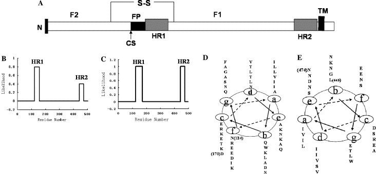
Heptad repeat regions of the APMV-2 F protein. (A) Schematic diagram of the APMV-2 F protein with the location of structurally significant domains. “S-S” represents the disulfide bond linking the F1 and F2; CS, cleavage site; FP, fusion peptide; HR, heptad repeat; and TM, transmembrane region. (B) HR1 and HR2 sequences were predicted by the ExPASy-Coils program. (C) HR1 and HR2 sequences were predicted by the LearnCoil-VMF program. (D) Helical wheel of the HR1 (amino acids 124–170) is depicted. (E) Helical wheel of the HR2 (amino acids 443–474) is depicted.