
Figure 2.
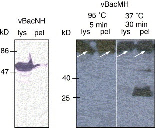
Western blot analysis of the lysates (lys) and pellets (pel) of Sf‐9 cells infected by vBacNH and vBacMH. The primary antibody was mouse anti‐His6 MAb while the secondary antibodies were either AP‐ (for rNH detection) or HRP‐conjugated (for rMH detection) goat anti‐mouse IgG. The membranes were developed with either BCIP/NBT color developing reagent or chemiluminescence reagent.