
Figure 4.
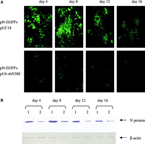
Silencing of N‐EGFP expression by siRNA in mouse muscles. Mice were injected with N‐EGFP expression plasmid and the siRNA expression vector in both tibialis anterior muscle. The N and EGFP expression was detected at day 4, 8, 12 and 16 post‐injection. (A) The muscles were isolated and frost slices were prepared, and EGFP expression was visualized under fluorescence microscope. The upper panel shows EGFP expression in mice injected with plasmid pN‐EGFP along with siRNA expression vector at various time post‐injection. The lower panel shows EGFP expression in mice injected with plasmid pN‐EGFP along with pUC18. (B) The muscles were isolated and N protein expression was detected by Western blot at different times. The upper panel indicates N protein expression in mouse muscles injected with plasmid pN‐EGFP along with: (1) pUC18 or (2) siRNA expression vector. The lower panel shows β‐actin expression as a loading control. The plasmid pUC18 used was to standardize the plasmid dosage for gene delivery in vivo. This figure shows the results of one representative experiment of two.