
Fig.2.
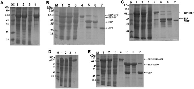
SDS–PAGE analysis of expression and purification of ELP-tagged protease and test proteins. Protein samples are separated by 12% SDS–PAGE and analyzed by Coomassie Brilliant Blue G-250 (20% methanol, 10% phosphoric acid, 10% ammonium sulfate, and 0.12% G-250) staining. (A) Expression and purification of ELP–3C protease. Lane M: molecular weight marker; lane 1: crude extract of cell expressing ELP–3C protease; lane 2: cleared lysate; lane 3: supernatant after precipitation of the ELP–3C protease; lane 4: purified ELP–3C protease. (B) Expression and purification of ELP–GFP and GFP. Lane M: marker; lane 1: crude extract of cell expressing ELP–GFP; lane 2: cleared lysate; lane 3: supernatant after precipitation of ELP–GFP; lane 4: purified ELP–GFP; lane 5: cleavage product of ELP–GFP; lane 6: precipitation of cleaved ELP, ELP–3C protease, and undigested ELP–GFP; lane 7: purified GFP. (C–E) Expression and purification of ELP–MBP (C), ELP-tagged SUMO protease (D), and ELP–SUMO–GFP (E). Gels are as described in panels (A) and (B).