
Figure 1.
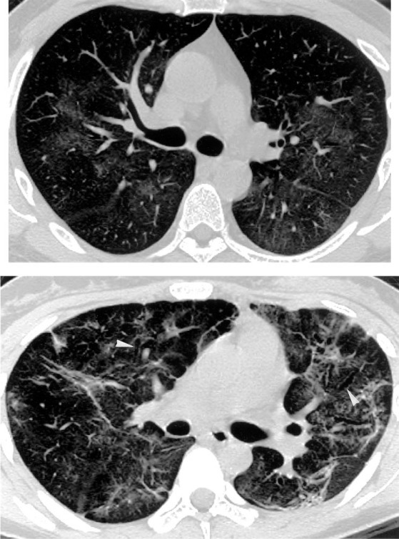
Top: HRCT scan of a 37-year-old man obtained 26 days after discharge from SARS-related hospitalization shows bilateral patchy areas of GGO without evidence of fibrosis, with random distribution in the transverse plane. Bottom: HRCT scan of a 22-year-old female SARS patient obtained 29 days after discharge shows random distribution of fibrosis consists of irregular linear opacities, traction bronchiectasis (arrowheads), and lung distortion. Concomitant presence of GGO is also visible.