
Figure 4.
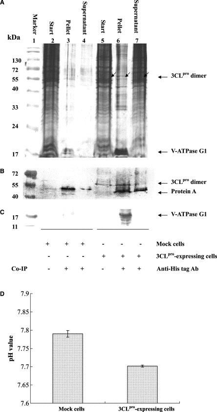
Cell‐based co‐immunoprecipitation (A–C) and decrease intracellular pH (D) by SARS‐CoV 3CLpro protease. The cell lysates of 3CLpro‐expressing cells and mock cells were immunoprecipitated by anti‐His tag to 3CLpro. The immunoprecipitate was analyzed using Coomassie blue staining (A), and Western blotting with anti‐His tag antibody to 3CLpro (B) and chicken anti‐V‐ATPase G1 polyclonal antibody (C). In addition, the 3CLpro‐expressing cells and mock cells were incubated with 20 μM carboxy SNARF‐1 for 30 min at room temperature. A dual‐emission ration at 580 and 640 nm of intracellular dye was detected using a fixed excitation at 514 nm, and then converted to intracellular pH (D).