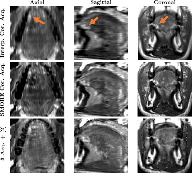
Figure 5: Comparison between SMORE(2D) and multi-view reconstruction for a tongue tumor subject:
Axial, Sagittal, and Coronal views of the tongue region in cubic b-spline interpolation and SMORE(2D) results for a single coronal acquisition, and multi-view reconstructed image [2] using three acquisitions. The arrows point out the bright looking scar tissue from a removed tumor.

