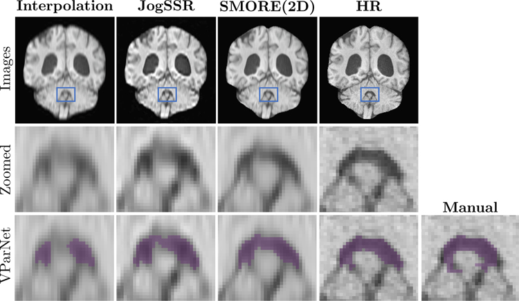
Figure 6: Coronal views of brain ventricle parcellation on an NPH subject:
The volumes with digital resolution of 0.8 × 0.8 × 0.8 mm that resolved from 0.8 × 0.8 × 3.856 mm LR image using cubic-bspline interpolation, JogSSR, SMORE(2D), and the interpolated 0.8 × 0.8 × 0.9 mm HR image. The patches in blue boxes are zoomed in the second row to show details of the 4th ventricle. The last row shows the VParNet [33] parcellation results and the manual labeling for the 4th ventricle.

