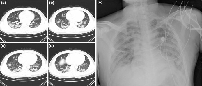
Extended Data Fig. 1. Chest radiographs of the patient.
a–d, Computed-tomography scans of the chest were obtained on the day of admission (day 6 after the onset of disease). Bilateral focal consolidation, lobar consolidation and patchy consolidation were clearly observed, especially in the lower lung. e, A chest radiograph was obtained on day 5 after admission (day 11 after the onset of disease). Bilateral diffuse patchy and fuzzy shadows were observed.

