
Figure 2. Structural changes between the pre- and postfusion F protein conformations.
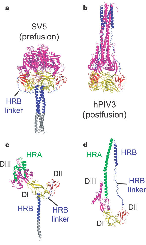
a, Ribbon diagram of the SV5 F-GCNt trimer. DI is yellow, DII is red, DIII is magenta, HRB is blue and GCNt is grey. b, Ribbon diagram of the hPIV3 (postfusion) trimer, similarly oriented by DI and coloured as in a. c, Ribbon diagram of a single subunit of the SV5 F-GCNt trimer, coloured as in a except for residues of HRA, which are green. d, Ribbon diagram of a single subunit of the hPIV3 F trimer, coloured as in c.