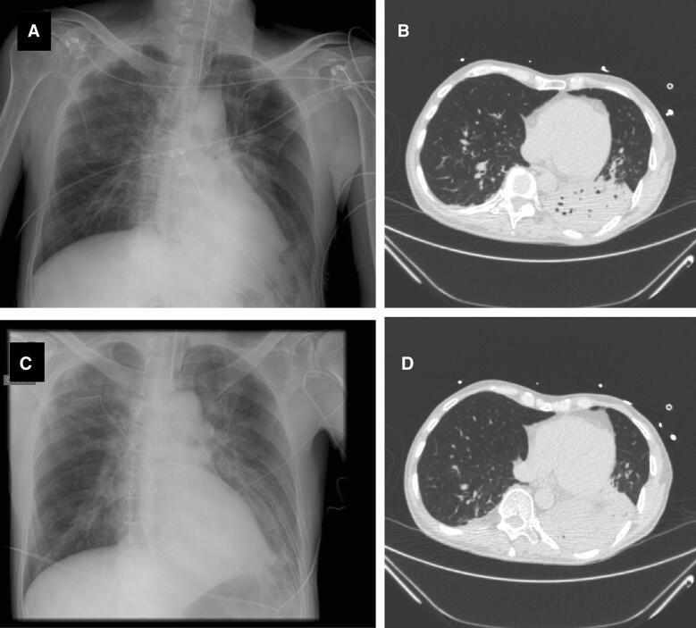
Fig. 1.
Chest X-rays and CT-scan of a 65-year-old man who developed ventilator-associated pneumonia. Chest X-ray performed the day VAP was suspected seems normal (a), whereas the CT-scan performed the same day showed consolidation of the left inferior lobe (b, d). Bronchoalveolar lavage yielded 105Enterobacter aerogenes. The next day, chest X-ray showed progression of pulmonary infiltrates (c). VAP diagnosis based on chest X-ray would have been delayed

