
Figure 2. Severity of lung histopathology in SCV-challenged macaques.
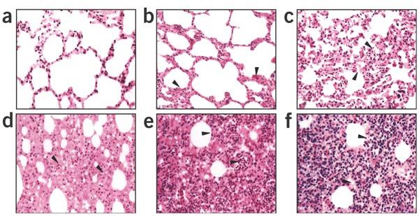
All lung histology sections were stained with H&E (original magnification, × 100). (a) Normal lung section from macaque #921 without SCV infection “−”. (b) Minor inflammation “±”, from macaque #138, slight broadening of alveolar septa and sparse monocyte infiltration. (c) Apparent inflammation “+”, from macaque #077, hemorrhage in septa, elastic fibers of alveolar wall distorted as shown by silver staining. (d) Early symptom of acute DAD “++”, from macaque # 015, alveolus septa broadening with increasing infiltration of inflammatory cells. (e) Typical symptom of acute DAD “+++”, from macaque #212, extensive exudation and septa broadening, shrinking of alveoli caused by pressure, restricted fusion of the thick septa, obvious septa hemorrhage, ruptured elastic fiber of alveolar wall and slight cell infiltration in alveolar cavities. (f) Severe acute DAD “++++”, from macaque #202, massive cell filtration and alveoli shrinking, sheets of septa fusion, necrotic lesions at the hemorrhagic septa and massive cell infiltration in alveolar cavities.