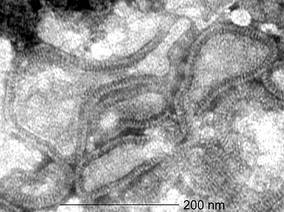
Figure 1. Electron micrograph of hMPV particles.

Virus concentrated from infected tMK–cell culture supernatants were visualized by negative contrast electron microscopy after PTA staining24. Magnification, ×92,000.

Virus concentrated from infected tMK–cell culture supernatants were visualized by negative contrast electron microscopy after PTA staining24. Magnification, ×92,000.