
Figure 3. Nucleotide and deduced amino-acid sequences for selected regions of the hMPV genome.
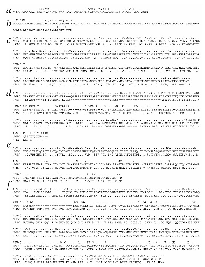
a and b, The nucleotide sequences for the 3′ end of the viral genome and the intergenic region between M and F ORFs. Note that the underlined sequence in a refers to the primer used for PCR amplification, and therefore does not necessarily reflect the actual hMPV leader sequence. c–f Comparison of the amino acid sequences of the putative N (panel c), P (panel d), M (panel e) and F (panel f) ORFs of hMPV, aligned with those of APV and RSV. Residues that differ between isolate 00-1 and the other viruses are shown, identical amino acids are represented by periods, gaps are represented by dashes.