
Figure 1. Histological lesions and immunohistochemical and ultrastructural detection of SCV in lungs of experimentally infected macaques.
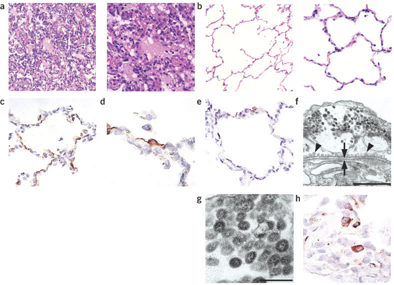
(a) DAD in pulmonary alveoli (control group). (b) Normal pulmonary alveoli (prophylactic group). (c,d) SCV antigen expression in type 1 pneumocytes (control group). (e) Lack of SCV antigen expression in pulmonary alveoli (prophylactic group). (f) Viral nucleocapsids in cytoplasmic vesicle of type 1 pneumocyte (control group), showing pinocytotic vesicles (arrowheads) and basement membrane (arrows). Scale bar, 1 μm. (g) Higher magnification of viral nucleocapsids shown in f. Scale bar, 200 nm. (h) SCV antigen expression in type 2 pneumocytes (control group). Sections were stained with H&E (a,b), for SCV antigen (c–e,h), or with uranyl acetate and lead citrate (f,g). Original magnification, ×25 (a left, b left), ×100 (a right, b right, c,e) or ×250 (d,h).