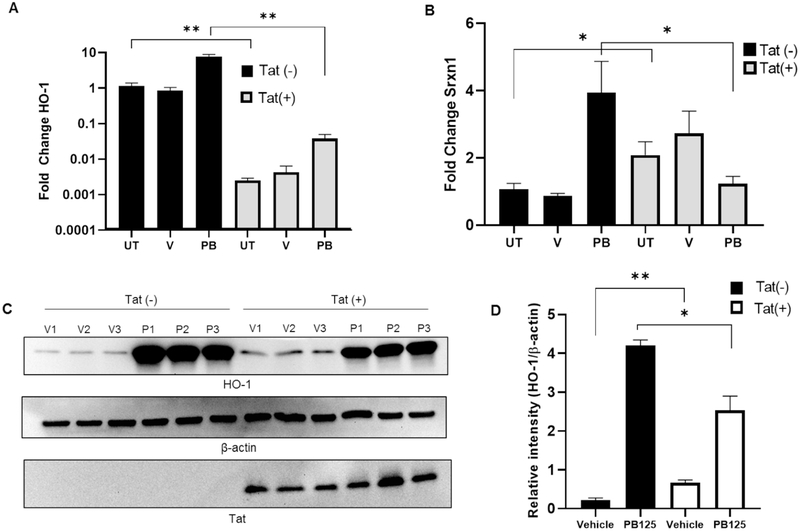
Figure 6. Effects of Tat on HO-1 and SRXN1 Expression in HPAEC.
(A) Tat represses HO-1 transcription in HPAEC. Cells were mock transfected or transfected with pTat-FLAG and incubated overnight with either regular growth medium or medium supplemented with vehicle (0.016% ethanol) or PB125 (8 mg/L). Total RNA was collected after overnight incubation and RT-qPCR was performed. Values shown as fold change relative to Tat(−) un-treated controls. Mean ± SEM of biological replicates (n = 6). (B) SRXN1 transcriptional induction is refractory to PB125 treatment in Tat expressing HPAEC. Experiment carried out as in A. Mean ± SEM of biological replicates (n = 6). (C-D) Induction of HO-1 Expression by PB125 is attenuated by Tat in HPAEC. Transfections and PB125 treatments were performed as in A. Total protein extracts were collected after overnight incubation for immunoblot analysis. Densitometry values expressed as relative intensity normalized to beta-actin. Mean ± SEM of biological replicates (n = 6). (**p ≤ 0.01 by unpaired student’s T-test) (UT = Un-treated, V = Vehicle, P/PB = PB125).

