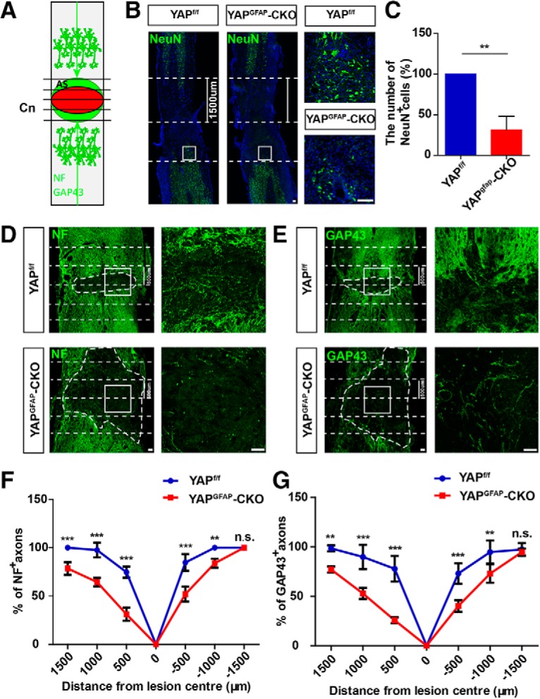
Figure 5.

Conditional yap deletion in astrocytes inhibited neural regeneration after SCI. A, Scheme of analysis of NF or GAP43 positive axons after SCI. Horizontal view of lesion core and astrocytic scar (AS) after SCI. Intercepts of NF and GAP43 positive axons with lines drawn at various distances from lesion center (Cn) were counted and expressed as a percentage of axons 1500 μm proximal. B, Immunostaining of NeuN (green) and DAPI (blue) in spinal cords of 2-month-old male yapf/f and yapGFAP-CKO mice at 14 d after SCI. C, Quantitative analysis of the number of NeuN-positive cells in the distance lesion center of the spinal cords as shown in B (n = 4 per group). D, E, Immunostaining of NF (D) and GAP43 (E) in spinal cords of 2-month-old male yapf/f and yapGFAP-CKO mice at 14 d after SCI. Dotted lines indicate lesion center 500 μm on either side. F, G, Quantitative analysis of NF positive (F) and GAP43 positive (G) area at various distances from the SCI lesion center as a percentage of the total area of axons present 1500 μm proximal (n = 5 per group). Dotted lines indicate lesion center and 1500 (B) or 500 μm (D,E) on either side. White dashed lines indicate the injury sites. B, D, E, Images of selected regions (white squares) are shown at higher magnification. C, Quantitative data were analyzed using Student's unpaired two-tailed t test. F, G, Data were analyzed using two-way ANOVA with Bonferroni post tests. **p < 0.01, ***p < 0.001, n.s., not significant. Data are mean ± SEM. Scale bars, 100 μm.
