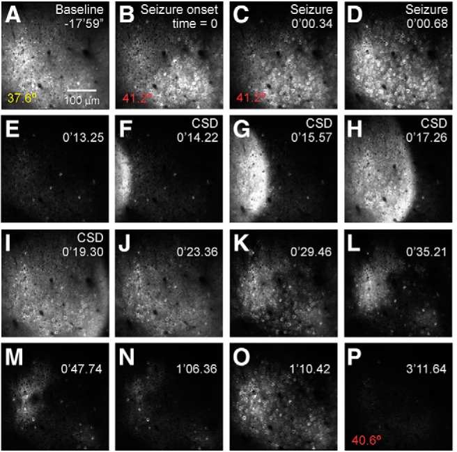
Figure 2.

In vivo 2P calcium imaging depicts the evolution of a naturalistic seizure at cellular resolution in an awake, behaving Scn1a+/− (DS) mouse. In this example, a mouse underwent passive warming to a core body temperature of 42°C while mobile on a spherical treadmill contained within a custom-made enclosure while head-fixed during ongoing imaging. Time during the imaging session relative to seizure onset is shown at the top right in minutes (′) and seconds (″), following a ∼15 min baseline recording period) and core body temperature (bottom left) is shown. A, Baseline GCaMP6f fluorescence imaged at 950 nm. B, Seizure onset, with hypersynchronous activation across the imaging field. C, D, Seizure propagation. E, Post-ictal silence. F–L, Cortical spreading depolarization. M–P, Inter-ictal discharges. Data were acquired at 29.4 Hz at 2× digital zoom using a 16× NA 0.8 water-immersion objective (Nikon). See Movie 1.
