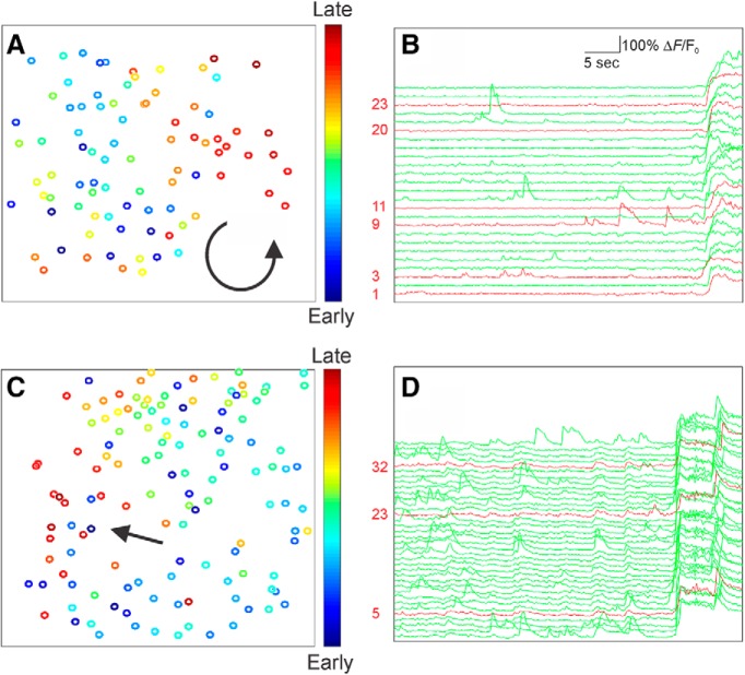
Figure 4.
Analysis of cellular-resolution imaging of seizure propagation shows widespread neuronal recruitment. A, C, Recruitment of identified neurons during two example seizures is color-coded, with early (cool colors) and late activation (hot colors). Direction of seizure propagation is illustrated with an arrow, for a seizure with a spiral pattern (A) and wave-like pattern (C). B, D, Selected individual ΔF/F0 traces for PV-INs (red) and putative excitatory cells (green) illustrating activity in the immediate preictal period.

