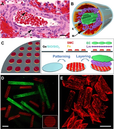
Fig. 1. Schematics and optical images showing biomimetic hSMPA fabrication and structural similarity to a human small muscular distal acinar pulmonary artery.

(A) Color photomicrograph of an adult hSMPA at the level of the respiratory bronchiole shown in cross section with hematoxylin and eosin staining. Black arrows indicate the EC of the intimal layer (E) and the VSMCs of the muscularis (M). Scale bar, 20 μm. Original magnification, ×200. This native artery exhibits several important structural characteristics including multicellular layering, curvature, and patterning. (B) Schematic illustration of the biomimetic hSMPA featuring patterning of cells and layering of VSMCs (M), laminin, and ECs (E). (C) Schematic illustration of the highly parallel, multistep patterning and assembly process for biomimetic hSMPA. Germanium (Ge) and bilayers of optically transparent silicon oxide and silicon dioxide (SiO/SiO2) were deposited on silicon wafers using electron-beam evaporation, followed by adhesive protein patterning and cell layering, which were all achieved in 2D. Upon dissolution of the sacrificial germanium layer in cell culture medium, the 2D bilayer films were released and then self-folded into tubes. Additional fabrication details are shown in the schematic in fig. S1, and snapshots of the roll-up process are shown in fig. S2. Fn, fibronectin; Lm, laminin. (D) Confocal microscope images of tubular constructs with tunable 1 and 2 mm length and protein pattern. Luminal surfaces of the tubular constructs were patterned with fluorescently labeled fibronectin (red) or bovine serum albumin (green). The distribution of protein fluorescence intensity is shown in fig. S8A. For cell culture, fibronectin without fluorescence labeling was used. Scale bar, 500 μm. (E) Epifluorescence images of rhodamine-phalloidin–labeled ECs growing on the luminal surfaces of biomimetic microvessels. Scale bar, 500 μm.
