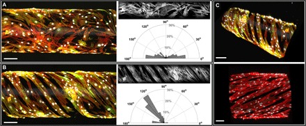
Fig. 5. Patterned fibronectin on tubular constructs oriented HPASMC adhesion.

(A) Confocal image (side view) of HPASMCs on an unpatterned tubular construct, stained for F-actin (phalloidin, red), smooth muscle α-actin (antibody labeling, green), and nuclei (DAPI, gray scale). The polar plot is based on image analysis of the gray scale F-actin image and shows that without patterning, HPASMCs attached and spread with random orientation. (B) Confocal image (side view) of HPASMCs grown on a patterned tubular construct, stained for F-actin (phalloidin, red), smooth muscle α-actin (antibody labeling, green), and nuclei (DAPI, gray scale). On the basis of image analysis of the gray scale F-actin image, the polar plot shows that F-actin filaments demonstrated alignment in parallel helical structures on the fibronectin-patterned scaffold. Binning in the polar plots is 10°. (C) 3D views of biomimetic microvessels demonstrating tunable variations in orientation angles and patterning periodicity, with labeled F-actin (phalloidin, red), smooth muscle α-actin (antibody labeling, green), and nuclei (DAPI, gray scale). Scale bars in (A to C), 100 μm.
