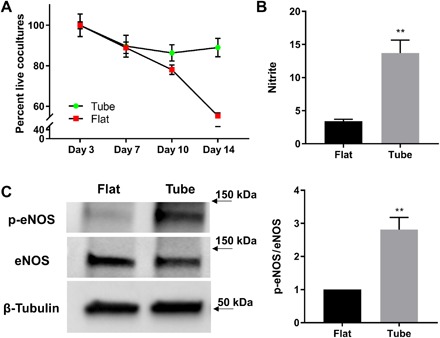
Fig. 6. Cellular longevity and cell signaling demonstrated markedly improved functionality in biomimetic hSMPAs.

(A) Cell viability was calculated as the percentage of live cells compared with baseline (day 3) using the CyQUANT assay. Coculture of HPMEC and HPASMC populations were assayed. Data from flat (red) and tubular constructs (green) were compared. Data are displayed as means ± SEM. (B) HPMECs in the biomimetic microvessels exhibited a fourfold rise in nitric oxide production over the levels observed in HPMEC monolayers in flat culture. Nitrite levels in cell culture medium were determined by using a Sievers bioluminescence nitric oxide analyzer. Nitrite levels were normalized to total protein content for each sample. The numbers in the y axis indicate picomoles of nitrite per micrograms of protein. Medium was collected 48 hours after confluency and medium replacement. Data are displayed as means ± SEM (**P < 0.01). (C) Phosphorylation of eNOS (at Ser1177) was found to be greater in HPMECs that were seeded and cultured on biomimetic microvessels than that in cells on flat SiO/SiO2 films. HPMECs were grown at each condition for 48 hours. Western blots of cell lysates from each population were analyzed with antibodies against phosphorylated eNOS (p-eNOS), total eNOS, and β-tubulin (protein loading control). Data are displayed as means ± SEM (**P < 0.01).
