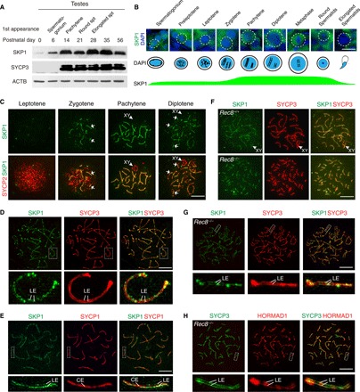
Fig. 1. Localization of SKP1 to SC LEs coincides with chromosome synapsis.

(A) Western blot analysis of SKP1 in developing mouse testes. Timing of the first appearance of spermatogonium, pachytene spermatocytes, round spermatids (spt), and elongated spermatids in developing testes is shown. SYCP3, meiosis-specific protein control; ACTB, loading control. (B) Spatiotemporal expression pattern of SKP1 in postnatal male germ cells. DAPI, 4′,6-diamidino-2-phenylindole. (C) Localization of SKP1 to synapsed regions of the SC (indicated by arrows) in wild-type (WT) spermatocytes. (D) Super-resolution localization of SKP1 and SYCP3 in pachytene spermatocytes. (E) Super-resolution localization of SKP1 and SYCP1 in pachytene spermatocytes. SYCP1 is a component of both CE and transverse filaments. (F) Localization of SKP1 to the SC between sister chromatids in Rec8−/− spermatocytes. (G) Super-resolution localization of SKP1 in Rec8−/− spermatocytes. (H) Super-resolution localization of HORMAD1 in Rec8−/− spermatocytes. Scale bars, 10 μm.
