
Fig. 1. RIBE is mainly mediated by RT-MPs.
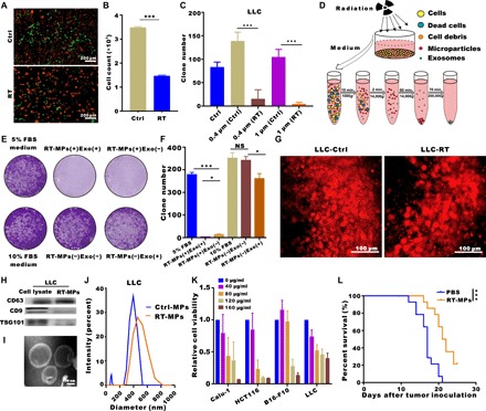
(A) Representative fluorescence microscope images of cell mixtures of unirradiated LLC-RFP and unirradiated LLC-GFP (top) and irradiated LLC-RFP and unirradiated LLC-GFP (bottom). Scale bars, 200 μm. (B) Flow cytometry analysis of green LLC-GFP cells in (A). (C) Quantification of clone formation after coculture with medium from irradiated cells using 0.4- and 1-μm pores. (D) Experimental outline for the production of microparticles and exosomes. (E and F) Representative images showing the colony numbers of LLC cells in the presence of microparticles or exosomes. NS, not significant. (G) Titanium chambers were surgically implanted in C57BL/6 mice, and irradiated or control tumor cells were then subcutaneously injected (2 × 10⁴) into the window. Irradiated tumor cells released more microparticles in the window chamber. Representative images are shown. Scale bars, 100 μm. (H) Western blots of CD9, CD63, and TSG101 expression in LLC whole-cell lysates (positive control) and RT-MPs pellets. (I) TEM images of RT-MPs. Scale bars, 100 nm. (J) Representative size and particle distribution plots of LLC-derived MPs and RT-MPs. (K) Calu-1, HCT116, B16-F10, and LLC cells were treated with various concentrations of RT-MPs for 48 hours, and cell viability was estimated using Cell Counting Kit-8 (CCK-8) assays. (L) Survival analysis in all two groups (n = 14 per group). *P < 0.05 and ***P < 0.001.