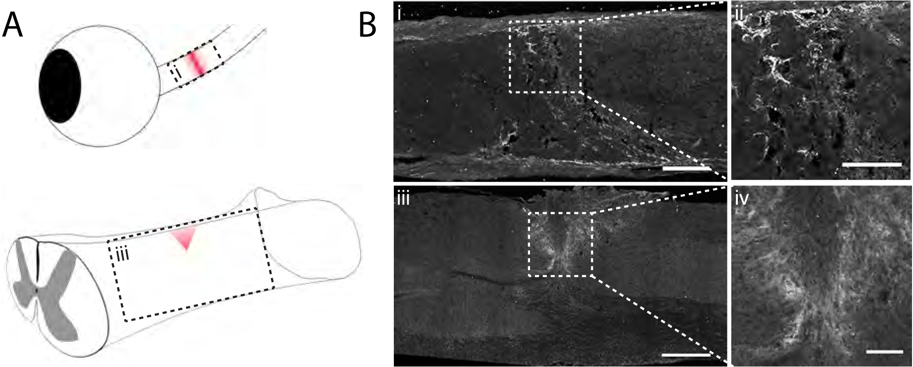
Fig. 1.

4-sulfated CSPGs accumulate in the glial scar after injury. A) Schematic diagram depicting optic nerve crush and dorsal column crush surgeries. Red shading indicates the area of the lesion following injury. B) Micrographs from the boxed regions in A showing injured mouse tissue 7 days after injury analyzed by immunohistochemistry with CS-56. Scale bar = (i) 100 μm, (ii) 50 μm, (iii) 400 μm, (iv) 100 μm
