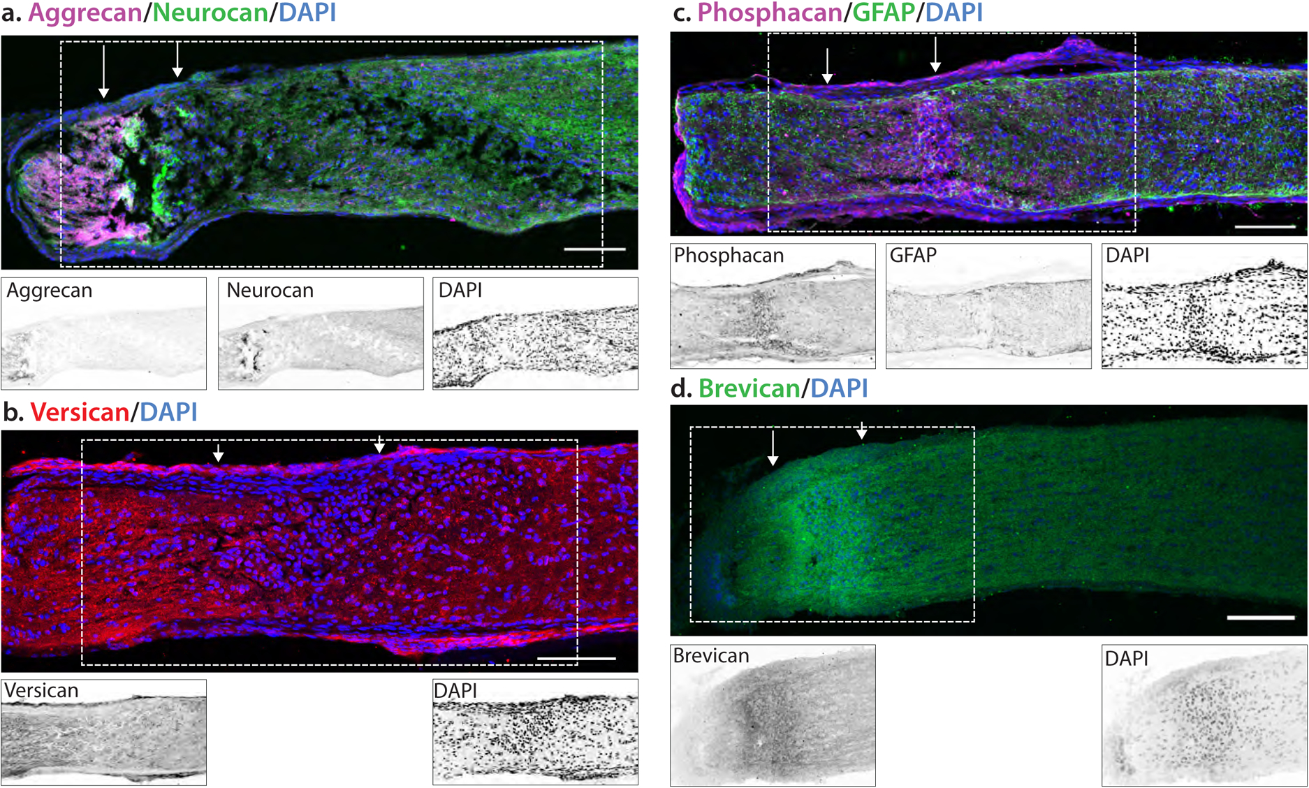
Fig. 7.

Localization of proteoglycan core proteins in 7 dpc injured mouse optic nerve. Mice were subject to a crush injury and tissues were obtained 7 days post crush. The localization of the crush is demarked by arrows. Insets are inverted images of the boxed area. (A) Aggrecan staining increased in the area proximal to the lesion, with low levels of staining in the lesion core devoid of GFAP. In contrast, neurocan staining was highest in the lesion core. (B) Versican levels were increased both proximal and distal to the lesion, with lower levels in the lesion core. (C) Phosphacan and (D) brevican staining were increased in the lesion core. Scale bar = 100 μm.
