FIGURE 2.
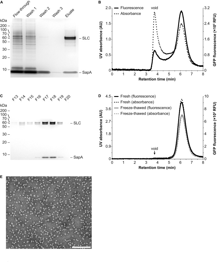
Purification and analysis of Salipro-SLC nanoparticles by chromatography. (A) Salipro nanoparticles containing SLC were separated by affinity chromatography from other Salipro-IMPs via the Strep-tag II affinity tag of SLC. Silver staining after SDS-PAGE of protein samples from the different purification steps showed that the eluate is pure as it contained only SLC and SapA. (B) Analytical SEC of the eluate from the affinity chromatography step with both UV (absorption at 280 nm) and fluorescence (excitation at 500 nm and emission at 512 nm) detection, able to monitor the daGFP reporter fused to SLC, revealed a homogenous population of Salipro-SLC nanoparticles. (C) The concentrated eluate from the affinity chromatography step was fractioned by preparative SEC. Fractions were visualized by silver staining after SDS-PAGE to verify the pure preparation of SLC in Salipro nanoparticles. (D) The pooled and concentrated SEC peak fractions [F17–19 in (C)] maintained their assembled state even after freeze-thawing as judged by analytical SEC. (E) Representative negative-stain electron micrograph showing a homogenous population of Salipro nanoparticles, which in this case each contain a single SLC. The scale bar represents 200 nm.

