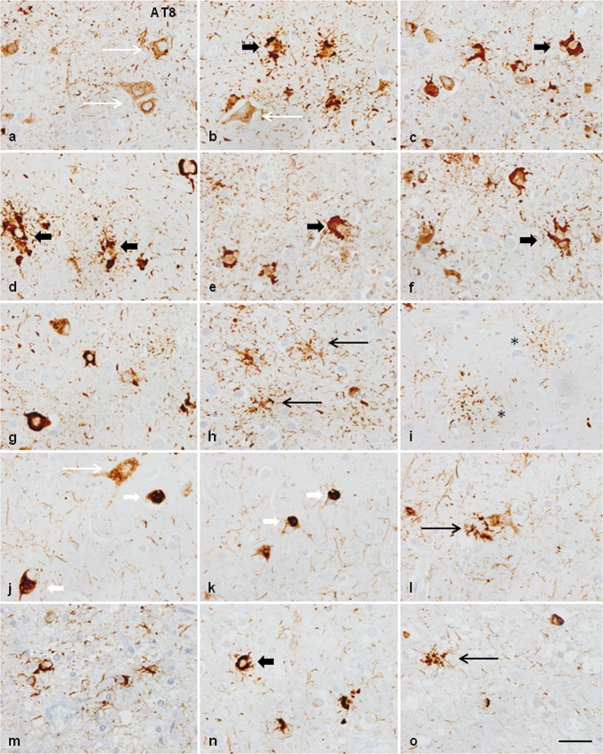
Fig. 2.
Characteristics of neuronal and astroglial phospho-tau deposits in the frontal cortex in GGT linked to MAPT P301T mutation; a–i: case 1; j–o: cases 3 and 4. In case 1, phospho-tau deposits, as revealed with the AT8 antibody, are finely granular in the cytoplasm, or form perinuclear halos, or globular tangles (long white arrow) (a, b, d, g). In cases 3 and 4, phospho-tau deposits are granular (long white arrow), globular, or round Pick-like bodies (short white arrow) (j, k). Astrocytic deposits in case 1 are typical GAIs with perikaryal globular structures or forming dense perinuclear inclusions of variable size consistent with immature stages (short black arrow) (d–f); together with astrocytes with longer cell and fine processes (long black arrow) (h), and astrocytic plaque-like structures (asterisk) (i). Astrocytic deposits in cases 3 and 4 show a predominance in astrocytes with longer cell processes (white arrows) (l, m, o) in addition to typical GAIs (short black arrows). Paraffin sections processed for AT8 immunohistochemistry and slightly counterstained with haematoxylin; bar = 25 μm

