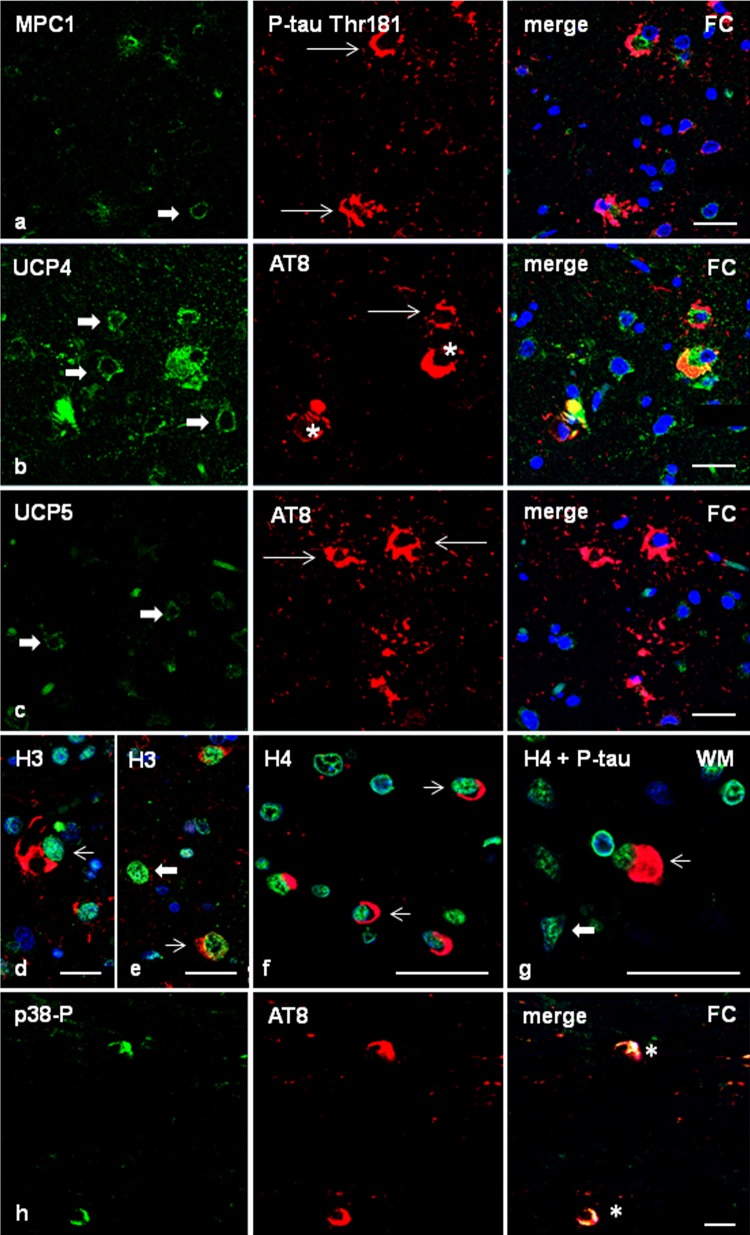
Fig. 5.
Frontal cortex in GGT linked to MAPT P301T mutation (case 1). Double-labelling immunofluorescence and confocal microcopy of the frontal cortex (FC) in case 1 using antibodies anti-MPC1 (a), UCP4 (b), or UCP5 (c) (green), and anti-P-tau Thr181 or AT8 (red). MPC1 is expressed equally in astrocytes with tau deposits (thin arrows) and without tau deposits (thick arrow) (a). Similarly, UCP4 is found in astrocytes bearing phosphorylated tau (thin arrows) and in cells without tau deposits (thick arrows) (b). In contrast, UCP5 is absent in GAIs deposits (thin arrows) when compared with cells without tau deposits (thick arrows) (c). Nuclei are counterstained with DRAQ5™ (blue). Paraffin sections, a–c, bar = 30 μm. d–g Expression of histones in frontal cortex and white matter in GGT (case 1). H3K9me2 immunoreactivity is expressed equally in cells with (thin arrows) and without (thick arrows) phosphorylated tau deposits (P-tauThr181 antibody) (d, e). Similarly, H4K12ac immunoreactivity is expressed equally in oligodendrocytes with coiled bodies (f) and globular inclusions (g) (AT8 antibody) (thin arrows), and in oligodendroglia cells without tau inclusions (thick arrows). Nuclei are counterstained with DRAQ5™ (blue). Paraffin sections, d bar = 20 μm; e–g bar = 30 μm. h phosphorylated p38 Thr180-Tyr182 (p38P) co-localizes with AT8-immunoreactive deposits (asterisk) in frontal cortex in GGT. Paraffin section without nuclear counterstaining, bar = 10 μm

