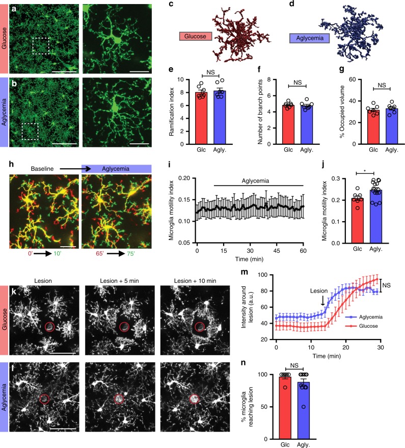
Fig. 3. Microglial surveillance is unaltered by aglycemia in situ.
a, b Microglial morphology following a 60-min incubation in glucose-containing aCSF (a, 10 mM glucose) or aglycemic aCSF (b, 0 mM glucose) (for panel a–g, glucose aCSF: n = 7 mice, 14 slices, 307 cells; aglycemia: n = 7 mice, 14 slices, 322 cells). c, d 3DMorph rendering of representative microglial cells following a 60-min incubation in glucose-containing aCSF (c) or aglycemic aCSF (d). e Ramification index of microglia 60 min after control or aglycemia incubation. f Number of branch points per microglia. g Percentage of brain volume occupied by microglia. h Process motility of microglia following one hour of aglycemia. Shown are overlaid images obtained 10 min apart to show dynamic microglial motility (for h–j, n = 7 slices for glucose, 15 slices for aglycemia). i Quantification of microglial motility index over time during aglycemia treatment. j Microglial motility index after 60-min incubations in glucose-containing aCSF or aglycemic aCSF. Shown here is the mean microglial motility for a 15-min imaging period following the incubation time (*p = 0.0319, unpaired, two-tailed t-test). k, l Microglial response to a laser-induced lesion following a 60-min incubation in glucose-containing aCSF (k) or aglycemic aCSF (l) (for k–n, n = 5 slices for glucose, 7 slices for aglycemia). m Quantification over time of the change in fluorescence intensity of the area around the lesion (not including the lesion). n Percentage of microglia surrounding the lesion (within a 75 µm radius of the lesion) with processes reaching the lesion within 20 min (n = 6 slices for glucose, 11 slices for aglycemia). For e–g, n, NS, p > 0.05, unpaired, two-tailed t-test; for m, treatment factor is NS, p > 0.05, but time factor is ****p < 0.0001, two-way ANOVA; data are represented as mean ± SEM. Scale bars, 100 μm (a, b, left panels); 20 μm (a, b, right panels); 25 μm h; 50 μm k, l. Source data are provided as a Source Data file. See also Supplementary Fig. 2.

