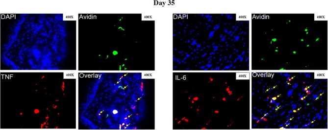
Figure 5.
Representative immunofluorescence staining of skin lesions from S. schenckii-infected WT mice at day 35. Representative immunofluorescence staining TNF (Red), IL-6 (Red), mast cells (Avidin, green), and DAPI (Blue, nuclei) of skin lesions from S. schenckii-infected WT mice at day 35. Red arrows: Double positive cells. Original magnification X40.

