Figure 2.
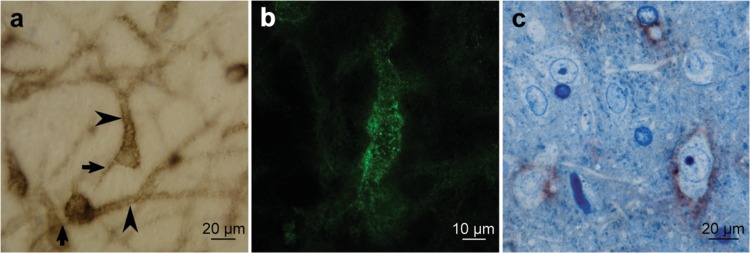
Golgi like stained cells in the mPOA of pregnant females are neurons. (a) Golgi like stained neurons in mPOA at GD18. Soma, dendrites (arrows), as well as axon initial segment (arrowheads) shows WFA associated DAB staining; (b) Confocal images z-stack of fluorescent labeled WFA preparations, showed that PNNs in the mPOA are organized in a “honeycomb” structure as described for neocortical PNNs together with a distributed punctuated pattern; (c) mPOA semithin sections at GD18 confirm the neuronal identity and show the external surrounding distribution of the WFA associated DAB staining. Positive surface stained cells show neuronal features, including big heterochromatic nuclei with prominent nucleolus, and abundant cytoplasm containing Nissl bodies. Interestingly, some neurons are not stained indicating that PNNs positive cells correspond to a neuronal subpopulation within this mPOA region.

