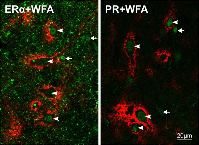
Figure 6.
PNNs associated neurons express estrogen receptor alpha and progesterone receptor. Double labeling using WFA and anti-estrogen-receptor-alpha antibody (ERα + WFA, left) or WFA and anti-progesterone-receptor antibody (PR + WFA, right). In both cases PNN-WFA label was tagged using streptavidin-594 (red), while hormone receptors were tagged using secondary antibodies conjugated with alexafluor-488 (green). In the left panel notice the presence of cell nucleus label for ERα and its variability between different PNNs expressing neurons (arrowheads). Positive nuclei belonging to PNNs negative cells (arrows) are also evident. Interestingly, there is a conspicuous doted label in the neuropilic region that is absent in neocortical neuropilic region (Supplementary Fig. 2). Note the presence of PR label nuclei in PNNs associated neurons in the right panel. PR label intensity is less variable compared to ERα label (left panel) and lacks the neuropilic dotted component. A population of PR labeled nucleus colocalizes with PNNs negative cells (arrows).

