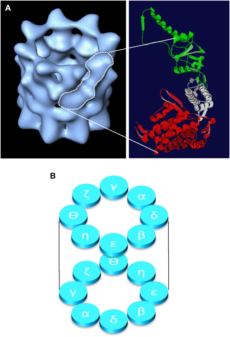
FIGURE 1.
Structure of the CCT oligomer. The structure of the CCT oligomer obtained by cryo-electron microscopy and three-dimensional reconstruction (Llorca et al., 2001) is shown in (A), left hand side with the approximate density of one CCT subunit outlined in white. The domain architecture of a single CCT subunit is shown in (A), right hand side based on the crystal structure of the alpha subunit of the thermosome (PDB 1A6D), where the equatorial domain (containing the nucleotide binding pocket) is shown in red, the flexible linker domain in gray, and the apical substrate binding domain is in green (adapted from Vallin and Grantham, 2019). The inter- and intra-ring subunit arrangements (Leitner et al., 2012; Kalisman et al., 2012) are shown in panel (B).

