
FIGURE 10.
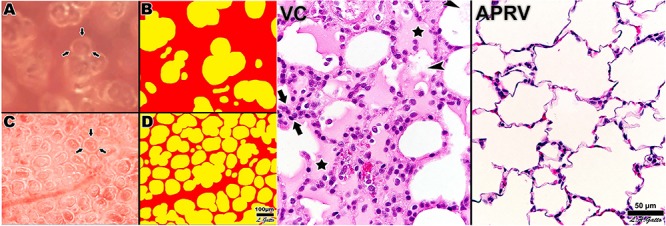
Left – Subpleural alveoli seen using in vivo microscopy in a rat hemorrhagic shock-induced ARDS model ventilated with volume cycled ventilation (VC; A,B) or airway pressure release ventilation (APRV) using the time controlled adaptive ventilation (TCAV) method (APRV; C,D). Individual alveoli are shown by arrows. Inflated alveoli were color coded yellow, and alveolar number, size, and surface area were measured by computer image analysis in each group. TCAV significantly improved alveolar patency and stability as compared with the VC group. Right – The APRV group delivered using the TCAV method stabilized alveoli that was associated with a significant reduction in lung histopathology as evidenced by open alveoli and reduced intra-alveolar edema (purple areas) as compared with the collapsed and edema-filled alveoli (stars), fibrinous deposits in the air compartment (arrowheads), and white cell infiltration (between arrows) in the VC group (Roy S.K. et al., 2013). Permissions obtained from Wolters Kluwer Health, Inc. License 4699411295724.