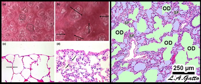
FIGURE 4.

Mechanical mechanisms of ventilator-induced lung injury (VILI) in the microenvironment: Left – In vivo subpleural alveoli in an acutely injured rat lung at inspiration (a) and expiration (b). Lung histology in the same rat lung injury model fixed at inspiration (c) and expiration (d) (Pavone et al., 2007). Alveolar recruitment/derecruitment (R/D) causing atelectrauma can be seen as fully recruited alveoli filling the microscopic field (white circles, two of which are highlighted with black dotted line) (a) that collapse during expiration (red atelectatic areas highlighted by arrows) (b). Histology shows open alveoli at inspiration with a shared alveolar wall highlighted by arrows (c) that collapse with expiration by alveolar wall folding, identified by arrows. Right – Stress-focus (SF) in areas with collapsed alveoli. SF adjacent to open alveolar ducts, causing alveolar duct overdistension (OD). SF-induced OD causes excessive mechanical stress and strain on alveolar duct walls, resulting in damage to pulmonary parenchymal cells (Ghadiali and Gaver, 2008). Loss of surfactant function results in alveolar instability (Figure 3, R/D). Histology at exhalation in a rat Tween-induced surfactant deactivation model with a tidal volume of 6 ml/kg and a PEEP of 5 cm H2O. Note the heterogeneous collapse of alveoli causing areas of stress-focus (SF). Adjacent to these areas of SF are overdistended (OD) alveolar ducts (Kollisch-Singule et al., 2014b). Summary – R/D causes excessive normal or “peeling” stress as the adhered alveolar walls peel apart during inflation; both R/D and SF cause OD in adjacent open stable tissue. This excessive stress and strain on alveolar walls results in lung parenchymal cell death and is a major VILI mechanism. Permissions obtained to reuse panels (a–d) (Pavone et al., 2007). Springer Nature license number 4766000130978.
