Fig. 2.
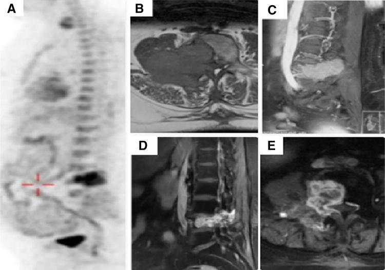
Metastatic carcinoma of papillary thyroid with spine metastasis at L4 vertebra. a FDG-PET showing solitary site of metastases. b, c Magnetic resonance imaging B (axial), c (sagittal) sections showing cord compression. d, e Follow-up images post radioactive iodine (RAI), angioembolization and radiotherapy

