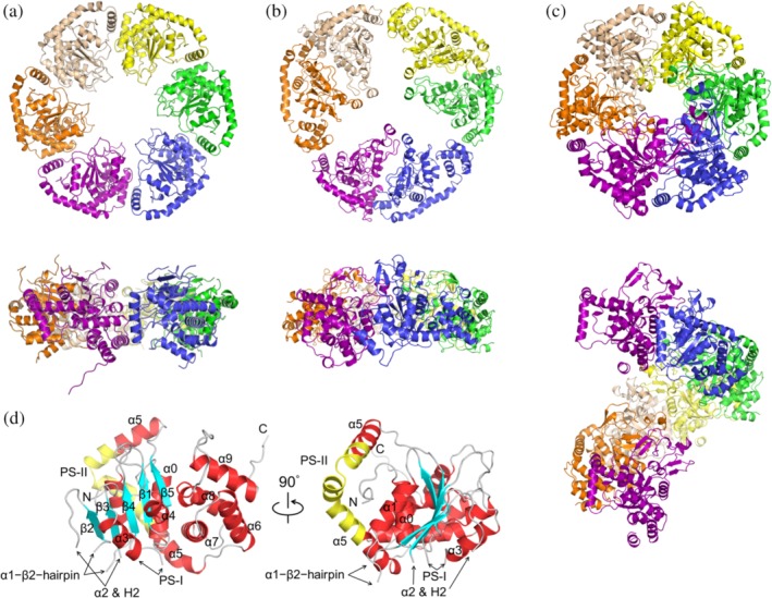
Figure 1.

Structure of the MgCh ATPase motor subunit in ribbon representation. (a) ChlI hexamer in top (upper panel) and side (lower panel) views. Six protomers (chains A–F) are colored in purple, orange, beige, yellow, green, and blue, respectively. (b) BchI hexamer with pseudo‐3‐fold symmetry (PDB: 2X31) reconstructed from the cryo‐EM maps. (c) BchI (PDB: 1G8P) in space group P65. The seventh chain along the c‐axis is shown in the same color as the first chain to present the rotational symmetry. (d) ChlI protomer (chain A). The α‐helices (red) and β‐strands (cyan) are labeled following previous conventions for AAA+ proteins.9, 16, 17 The three β‐hairpins, α1–β2–β‐hairpin, H2‐insert (within α2), and PS‐I insert (presensor I insert, between α3 and β4) are denoted by arrows; the PS‐II insert (presensor II insert, within α5) is in yellow; the loops are in gray
