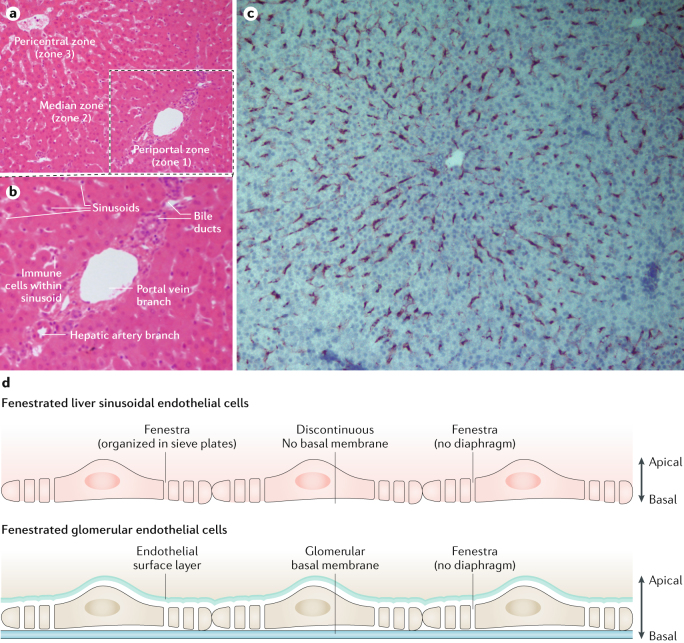
Fig. 1. Microanatomy of the human liver vascular tree.
a | Low-power image of human liver tissue (stained with haematoxylin and eosin) illustrating the lobular organization of the liver, with zonal architecture indicated relative to the position of the portal tract. b | Expanded periportal section of the same image to illustrate the different vascular compartments within the parenchyma. c | Immunohistochemical staining of stabilin 1, which highlights liver endothelial cell distribution within hepatic tissue in a normal liver section. d | A comparison of the structure of liver sinusoidal endothelium and glomerular endothelium.

