
Fig. 5. SRSF7-GFP promotes the formation of higher-order assemblies in vitro.
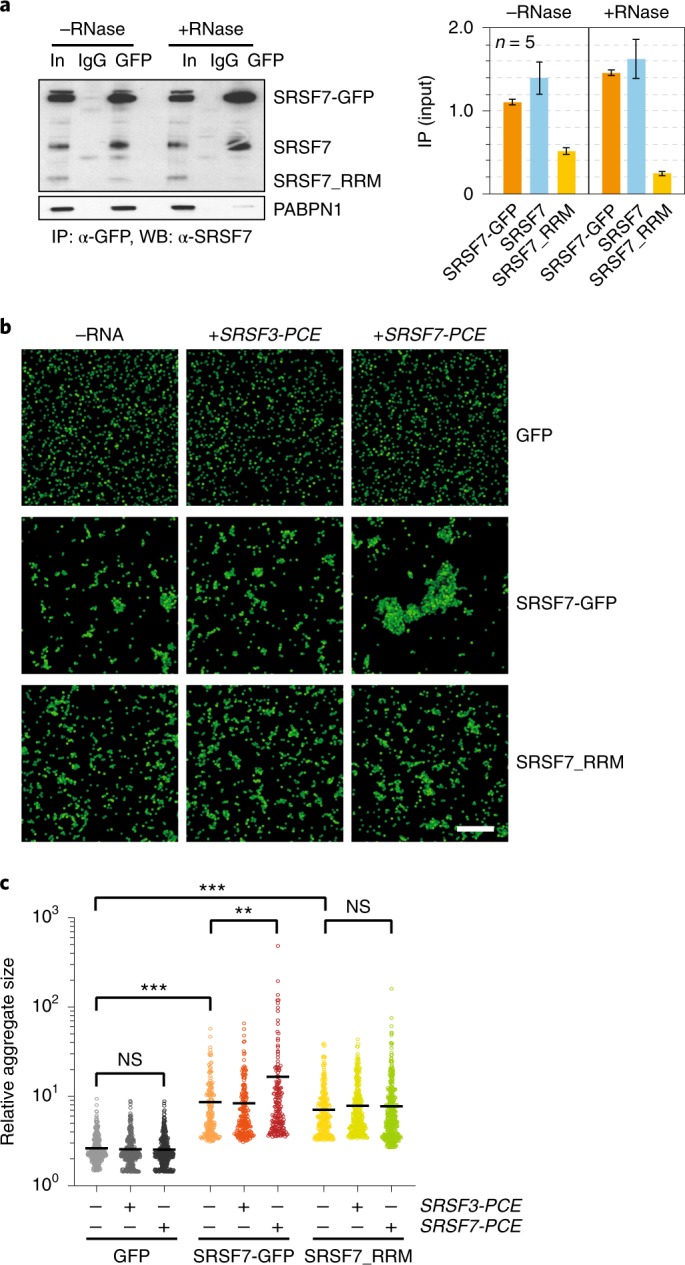
a, Left: co-IP of purified SRSF7-GFP probed with α-SRSF7 to verify the interaction with endogenous SRSF7 and SRSF7_RRM. PABPN1 was used to control for RNase treatment. Right: quantification of five Co-IP experiments with and without RNase treatment. Mean and error bars, s.e.m.; n = 5 independent experiments. b, SRSF7-GFP and SRSF7-PCE transcripts promote the formation of higher-order assemblies in vitro. Bead aggregation assays were performed with magnetic beads coupled to α-GFP antibodies that immunoprecipitated GFP, SRSF7-GFP or SRSF7_RRM-GFP from P19 cell lysates. Their aggregation propensity was tested in the absence or presence of transcripts corresponding to the PCE of SRSF7 or SRSF3. Shown is a representative confocal micrograph of fluorescent single beads and aggregates at ×40 magnification from three independent experiments. Scale bar, 50 µm. c, Quantification of beads per aggregate using FIJI (Analyze particles). Shown is the top 25th percentile of aggregate areas normalized to the area of one bead. This experiment is representative of three independent experiments. Center, median. ***P < 0.0002; **P < 0.002 (two-tailed Mann–Whitney test); NS, not significant. Uncropped blot images for a and data for graphs in a and c are available as source data.