
Figure 3. Diagram of the structure of the HIV-1 envelope glycoprotein gp41.
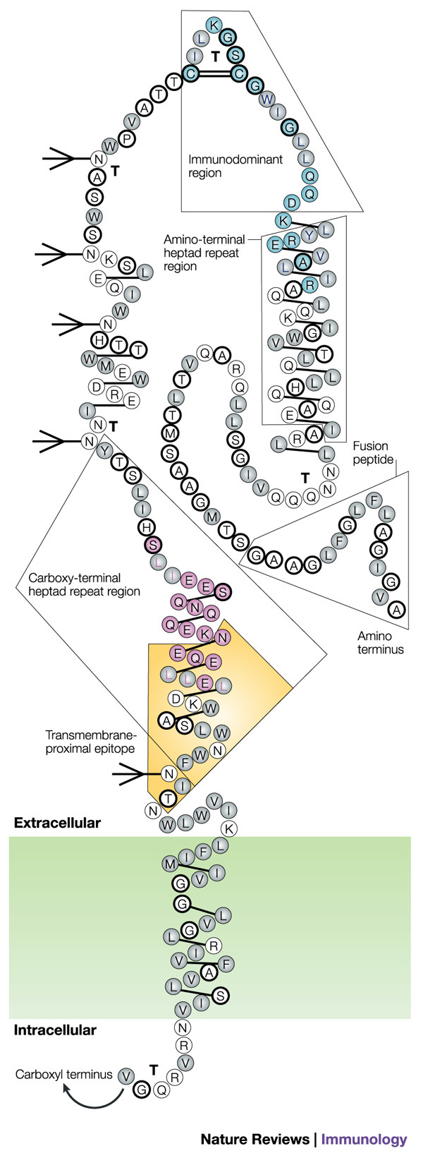
The gp41 molecule with α-helices depicted as alternating three- and four-amino-acid groupings connected by single lines, hydrophobic amino acids indicated as grey circles, charged amino acids as unfilled circles and neutral amino acids as heavily outlined circles. Strong turns are indicated by 'T', and potential glycosylation sites by branched-stick figures. Key functional and structural regions are boxed, and well-characterized epitopes are shown in colour, with epitope clusters I and II shown in turquoise and pink, respectively, and the transmembrane-proximal epitope highlighted in yellow. Reproduced with permission from Ref. 126 © (1989) Mary Ann Liebert Inc. Publishers.