
Figure 3. Model of a viroporin promoting viral budding at the plasma membrane.
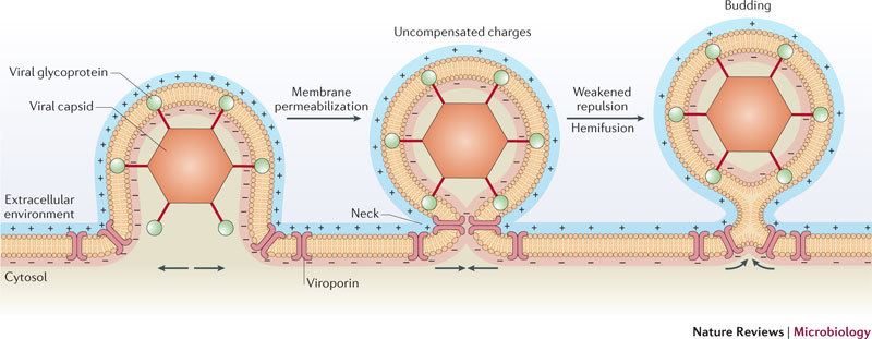
Viroporins localize at the plasma membrane in specific sites surrounding the neck of the budding virus particle, as described for the influenza A virus matrix protein 2 (M2) viroporin80. Viroporins alter membrane permeability by conducting the flux of different ions (for example, Na+ and K+) across the membrane in favour of their concentration gradients and so reducing the transmembrane potential, which is essentially determined by three factors: the concentration of ions inside and outside the cell; the permeability of the cell membrane to those ions (that is, the ion conductance) through specific ion channels; and the activity of electrogenic pumps (for example, the (Na++K+)ATPase and Ca2+ transport pumps) that require energy to maintain the ion gradients across the membrane. Depolarization of the membrane (that is, decreasing the imbalance of charges across the membrane) leads to a reduction in the charge density on the membrane surface. This phenomenon would result in a decrease in the electrical or contact repulsion between opposing monolayers of the membrane at the neck of the budding site (middle) and could provide the stimulus and even the energy to locally promote budding and release21,75, as occurs in depolarization-dependent exocytosis76.