
Figure 2. Inhibition of VHSV in cell cultures secreting ScAb.
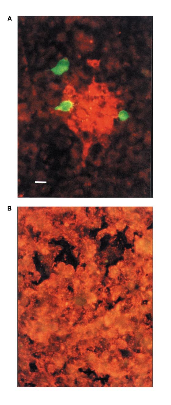
Cultures of fish cells were transfected with pcDNA3-BU1 (A) or pcDNA3 plasmid DNA (B) before inoculation with VHSV. Three days after inoculation the cultures were fixed and stained by dual-color immunofluorescence. Cells expressing ScAb were detected by using FITC-conjugated antibody (green fluorescence). Virus-infected cells were detected by TRITC-conjugated antibody (orange-red fluorescence). Bar corresponds to 10 μm.