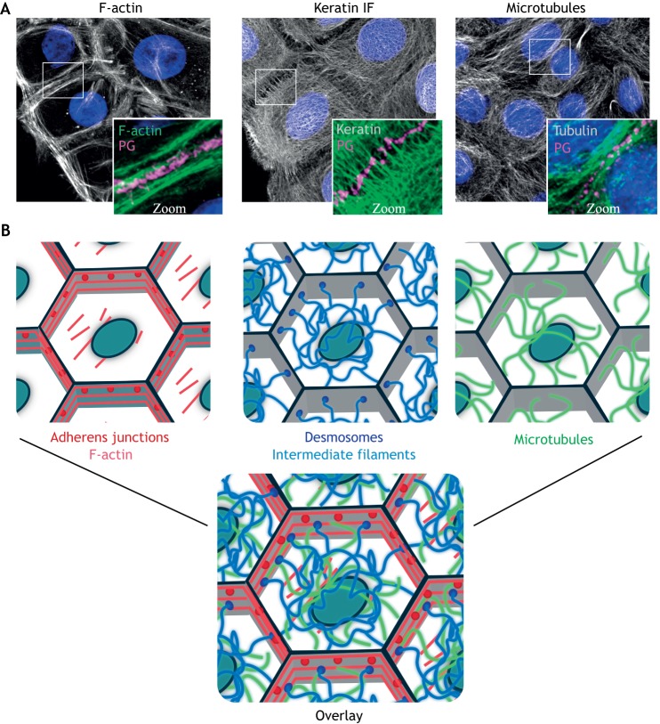
Fig. 1.
Architecture of the three main cytoskeletal systems. (A) Immunofluorescence staining shows the organization of the F-actin, keratin IF and microtubule cytoskeletons in human epidermal keratinocytes. Plakoglobin (PG) is used to show regions of cell–cell contact and nuclei are shown in blue with DAPI. (B) Schematic representations of the indicated filamentous cytoskeletons and associated cell–cell adhesive complexes, as well as an overlay to illustrate their interconnected nature. Gray outlines represent cell–cell junctional areas and ovals represent nuclei. Images supplied by the laboratory of K.J.G.

