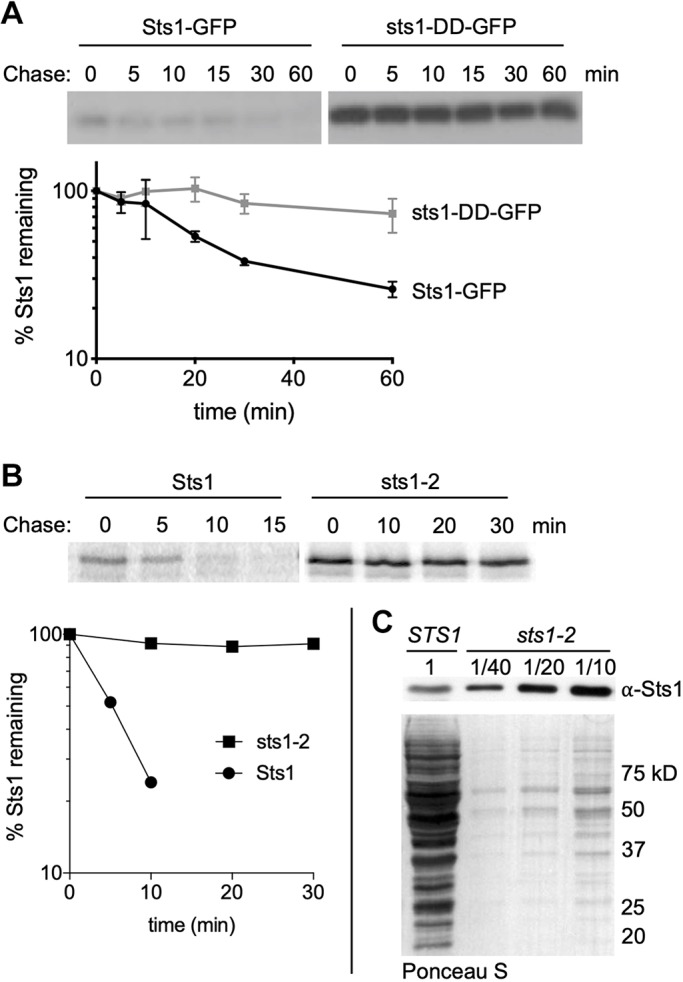
Fig. 3.

Degradation of wild-type and mutant forms of Sts1 in cells. (A) Cycloheximide-chase analysis was performed to determine the degradation rates of Sts1-GFP and sts1-DD-GFP using anti-GFP immunoblotting. MHY500 cells carried either p415MET25-STS1-GFP or p415MET25-sts1-DD-GFP and were grown at 30°C; cycloheximide was added at time 0 to block further protein synthesis. For quantitation, Sts1-GFP and sts1-DD-GFP levels were normalized to a PGK loading control. (B) Radioactive pulse-chase analysis of Sts1 degradation in MHY9692 (STS1) and MHY9693 (sts1-2) cells at 30°C. Proteins were immunoprecipitated using affinity-purified anti-Sts1 antibodies. Bottom panel shows the phosphorimager quantification of degradation rates. (C) Cell extracts from MHY9692 and MHY9693 were separated by SDS-PAGE and immunoblotted with anti-Sts1 antibodies. Extracts from MHY9693 were diluted to the indicated fraction of extract loaded for MHY9692. Ponceau S-stained membrane shows relative amounts of total protein extract.
